Imaging in Medicine (IMG)
Are you curious how cutting-edge imaging technologies reveal the inner workings of the human body and brain? In the Imaging in Medicine specialization, you’ll explore the physics and simulation of clinical systems such as Positron Emission Tomography (PET), Magnetic Resonance Imaging (MRI), and Computed Tomography (CT), while mastering image reconstruction, AI-driven processing, and brain data visualization. If you want to combine physical principles, high-performance computing, and medical innovation to better understand diagnostics and the human connectome, this specialization really matches your interests!
Description of the Specialization
The first part the lectures will cover the basics of imaging in medicine and biology, particularly Tomographic Imaging. There will be a focus on imaging with Ionising Radiation X-ray, CT, SPECT, PET. The related physics basics will be introduced and their relevance for imaging will be discussed. Computer-based simulation will be introduced to compare different physical phenomena. The lectures will present the concept and physical background of various devices applied during imaging starting from the data acquisition till the reconstruction of tomographic images. The complete processing pipeline will be presented and explained in detail. In the second part of this course the basics of image processing in general and image analysis of medical images in particular will be presented in all details. The application of image analysis in medicine and biology, starting from the basic description and basic operations on images extending to image registration and image fusion, will be demonstrated.
Module 1. Imaging 1 (IMG1) – mandatory
Workload: 8 ECTS (240 hours, 1 semester)
Final assessment: 30-minutes oral exam, not restricted in attempts
Components:
• Quantitative Medical Imaging (IMG1-a)
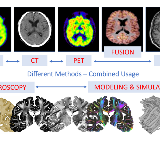
- Introduction to the Most Relevant Clinical Medical Imaging Devices: PET, CT and MRI
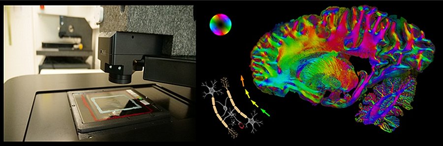
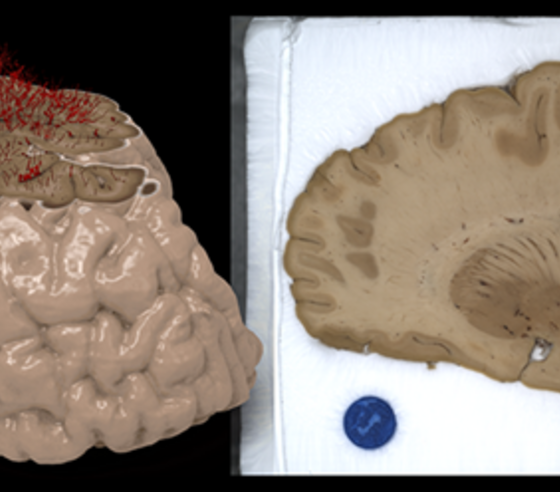
- Introduction to Microscopic Techniques Used for Microstructural Medical Imaging
- Physical Effects Used to Generate Contrasts (e.g., Decay, Absorption, Spin Dynamics)
- Image Reconstruction and Interpretation for Diagnostics
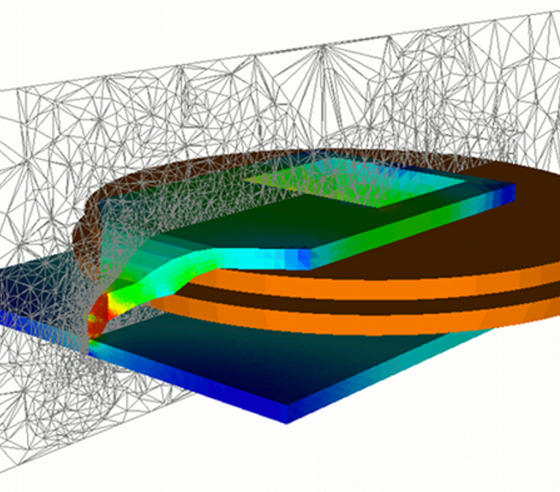
- Combined Usage of Experiment and Simulation
- Modelling of Phantoms and Simulation of Imaging Devices and Measurements
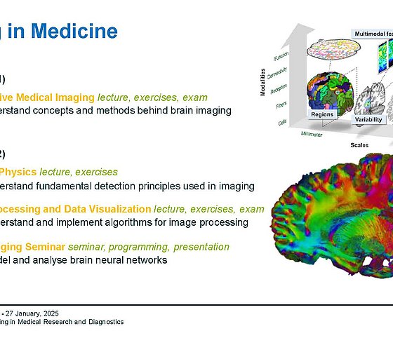
- Revealing the Human Connectome
• Detector Physics (TPDP-a)
Linear and Circular Accelerators. Betatron and Synchrotron Oscillations. Linear Beam Dynamics. Interaction of Particles With Matter, Shower, Momentum- and Track- Measurement, Track Detectors (Gas Chambers), Semi-Conductor Detectors, Time Measurement, Energy Measurement (Calorimeter), Particle Identification, Experiments of Particle and Astroparticle Physics, Instrumentation, Data Acquisition. Upload of protocols of two experiments in sufficient quality is needed to complete.
• Exercises Detector Physics (TPDP-b)
Contents of the lecture are practiced in dedicated exercises. Upload of at least 10 solutions of the exercises corresponding to at least 50% of the points is required to complete.
Module 2. Imaging 2 (IMG2) – mandatory
Workload: 8 ECTS (240 hours, 1 semester)
Final assessment: oral or written exam, not restricted in attempts
Components:
• Image Processing and Data Visualization (IMG2-a)
- Introduction to the Importance of Modern Image Processing and Data Visualization Techniques to Brain Imaging
- Data Types and Structures (Scalar, Vector, Volume Data)
- Transformation and Filtering Techniques to Carve Out Specific Image Features
- Image Processing Pipelines in a Supercomputing Environment
- Impact of AI on Image Processing
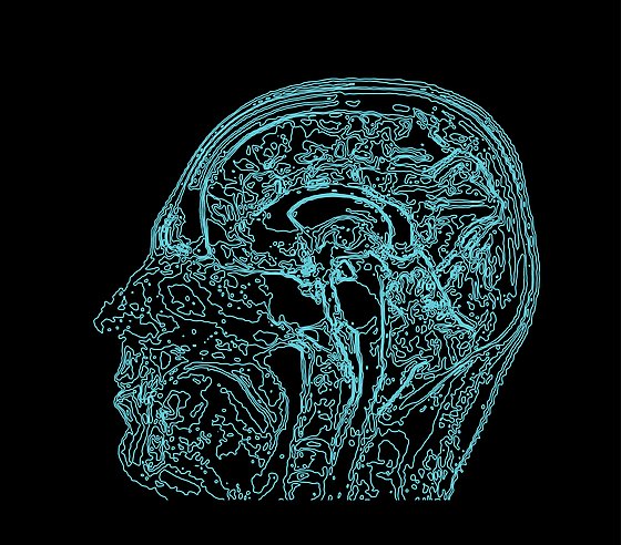
- Methods for Brain Data Visualization
• Seminar on Imaging II in Jülich (IMG2-b)
- Introduction to a selected modelling and simulation framework (lectures, online descriptions)
- Getting familiar with the code and its application (task-driven)
- Preparation of oral presentation about experience, method and results
The ungraded achievement (exercises, term paper, etc.) is required to complete.
At least 24 ECTS credits (or 13% of completed Bachelor´s degree) in the following fields: Atomic Physics, Nuclear Physics, Basic Quantum Mechanics. Good knowledge in Atomic and Nuclear Physics will be beneficial for the first part. Basic experiences in using computer-based analysis programs like ROOT, Matlab, Fiji may be advantageous for the second part of lectures.
You can check yourself, if you can study on this specialization by completing the:
Graduates of this specialization are qualified for careers in medical technology, biomedical engineering, healthcare industry, and research institutions. They find employment in companies developing and optimizing imaging systems such as X-ray, CT, SPECT, and PET, as well as in hospitals, clinical research centres, and medical software companies. Typical positions include Medical Imaging Engineer, Imaging Scientist, Image Processing Specialist, Clinical Application Engineer, R&D Engineer for diagnostic systems, and Medical Data Analyst. Alumni work on tomographic image reconstruction, simulation of imaging processes, development of image processing and registration algorithms, image fusion techniques, and optimization of diagnostic workflows. The specialization also provides strong preparation for doctoral studies and research careers in medical physics, biomedical imaging, and computational image analysis.
TBA
Contacts
Person responsible for the specialization:
Prof. Dr. Markus Axer, +49 202 439 3523, maxer[at]uni-wuppertal.de
Lecturers:
- Prof. Dr. Markus Axer (maxer[at]uni-wuppertal.de) – Image Processing and Data Visualization
Links:
Website of the working group on Fiber Architecture
Prof. Axer´s webpage on the website of the Research Center Jülich
Last modified: 25.03.2026